By Jay Kang
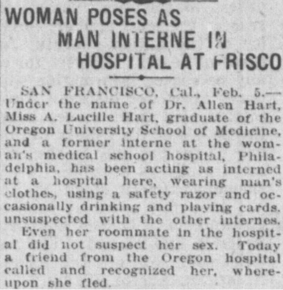
In February 1918, the Austin American ran the headline “Woman Poses as Man Interne in Hospital At Frisco.” (Figure 1). A bright young man had just applied for a position at a San Francsico hospital as a medical intern when a former colleague from Oregon recognized him – except, he previously knew this man as a woman. This information eventually reached the hospital superintendent, resulting in the young man in question fleeing California and returning home to Oregon. This young man was Alan L. Hart, and he would soon revolutionize tuberculosis care with the use of X-ray diagnostics.
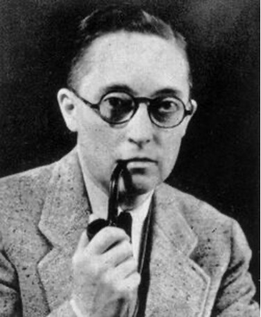
Alan Hart enrolled at the University of Oregon Medical School in 1913 and graduated as head of the class. Though in medical school Alan identified as a woman, he realized that he was happiest when he identified as a man. During his time in medical school, prior to his transition, he became the first person “assigned female at birth” to earn the coveted Saylor medal for being the top scholar in each of the school’s departments. His colleagues held high regards for Alan; they all complimented him for his brilliance and thought the award was rightfully given. Regarding his transition, he became one of the first trans men to receive a hysterectomy, a surgical procedure to remove the uterus. In addition to surgery, Alan also cut his hair short, dressed in masculine clothing, and started his career as a practicing male physician.
After being outed as a trans man in San Francisco (Figure 1), Alan attempted to resume his practice in Oregon, but his previous identity continued to haunt him. Everywhere he went, it seemed he could not escape being “outed” or having his biological sex be released to his patients. He would move to at least seven more states before eventually being accepted to University of Pennsylvania’s Master’s program in radiology in the 1920s. In academia, at least, his scholarly merit might be viewed as more important than his past. X-rays were invented in 1895, making this radiology technology new and impressive. At first X-rays were used to locate obstructions such as bullets or fractures in the bone; Dr. Hart would later use this technology to identify signs of a terrible disease before the onset of symptoms.
During this time, The United States was undergoing a massive public health crisis: tuberculosis (TB) was ravaging the country. TB is caused by an uncontrolled infection with Mycobacterium tuberculosis bacterium (MTB)1. MTB is a pathogen that infects the lungs and subsequently causes a number of symptoms such as fever, tiredness, and cough. Shockingly, TB claimed one in seven of all human lives during the 1800s.
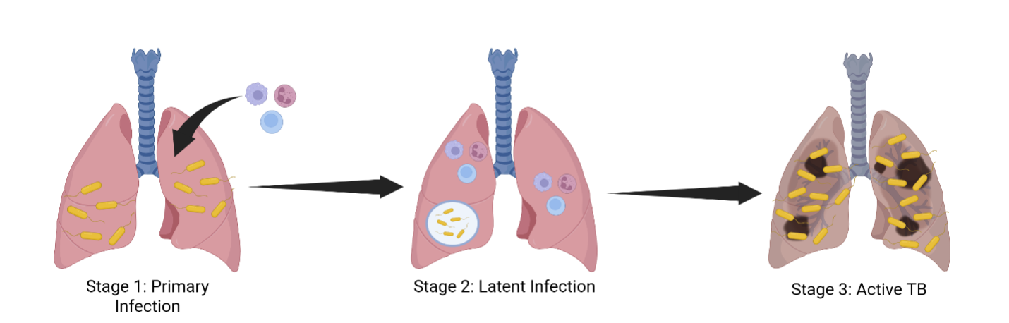
An MTB infection can be classified in three stages: primary infection, latent infection, and active TB disease. When someone is first infected with MTB, they will experience a primary TB infection, in which the host’s immune system is able to identify and contain the germs causing the infection within the lungs. However, this bacterium is known to be particularly resilient and often can escape the immune system and continue to multiply. One way MTB escapes immune-mediated control is by infecting macrophages. Macrophages are a part of the first line of defense against MTB; they phagocytose, or engulf and kill, invading pathogens. MTB hijacks macrophages, forcing them to initiate their own cellular death 2,3. As TB disease progresses past primary infection, the immune system is able to gain some control over the pathogen. By continuously fighting against MTB activity, it prevents the patient from experiencing any profound symptoms and MTB will eventually become latent. However, should the immune system fail to keep up with the infection, the bacteria will multiply, and the host will experience active TB disease (Figure 2).
During active TB cases, patients experience a cough that lasts for 3 weeks or longer, chest pain, fatigue, and weight loss. TB has the ability to extend past the lungs – a phenomenon known as extrapulmonary tuberculosis – and can additionally cause damage in areas like the voice box, kidneys, liver, and lymph nodes. Approximately half of all patients with active TB that go untreated will eventually die.
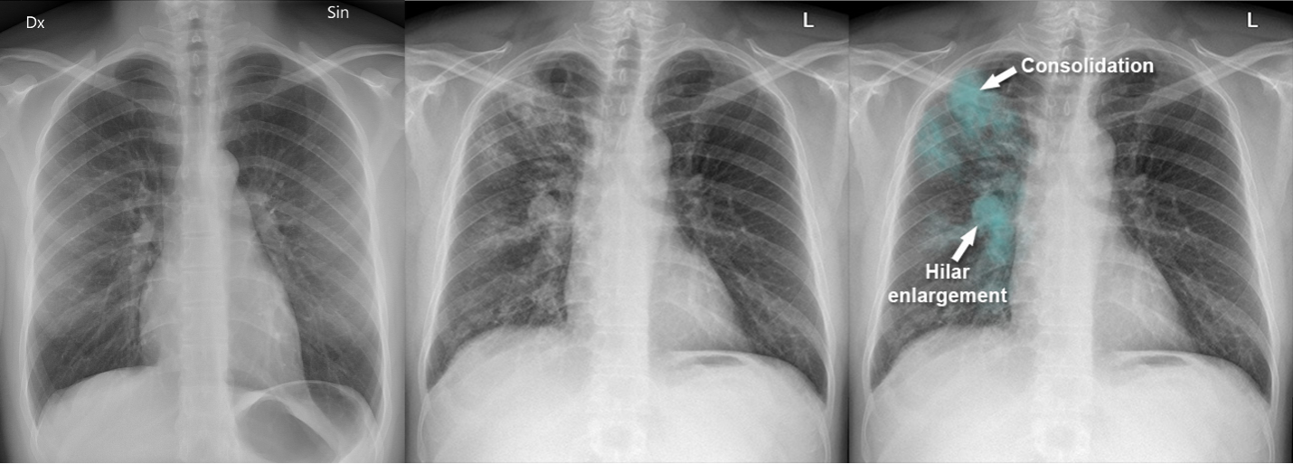
One of the key reasons why TB is so uncommon in the modern era is due to advanced technology that is able to detect MTB prior to the onset of active TB disease. Latent TB infection can remain dormant in the body for years, and sometimes even decades4. So – how did doctors figure out how to detect the disease before it entered the active TB phase, and thus prevent TB-mediated death? Dr. Alan Hart, interested in helping this particularly vulnerable population during a time when TB was one of leading causes of death, devised a way to utilize chest x-rays for early detection of MTB in a patient’s lungs (Figure 3).
Prior to Hart’s work, X-rays were primarily used after the onset of serious TB symptoms, rather than as a preventative measure5; no one had thought of using x-rays to get ahead of the disease. In doing so, Hart and many other doctors were able to help patients get the treatment they needed before the disease advanced, and isolate contagious patients to prevent spread to the general public6,7.
While chest x-rays alone are unable to confirm that a person specifically has TB because an abnormality could be evident of other pulmonary diseases, they are still powerful diagnostic tools that help clinicians identify mild symptoms as something potentially much more serious. Chest abnormalities can include consolidation, which are areas of the x-ray that present cloudy due to build up of fluids like blood or solid materials like fat. Additionally, an x-ray may reveal a hilar enlargement – the hilar is a region of the lung containing bronchi, blood vessels, and nerves. X-rays, combined with more modern preventative measures such as a blood or skin test, have helped prevent TB from consuming additional lives, sometimes doubling the rate of decline of TB infections. X-rays were so effective that by the time antibiotics were introduced to treat TB, TB deaths were reduced to 1/50th. In a different metric, TB deaths in the United States decreased from 194 deaths per 100,000 people in 1900 to 40 deaths per 100,000 people.
Today, chest X-rays are a cheap but effective tool, useful for eradicating TB globally. You may have noticed that you almost never hear about TB outbreaks in public. This decline is due to the advancement in detection and treatment of TB. The World Health Organization (WHO) aims to reduce TB incidence by 80% and reduce deaths by 90% by 2030. To do so, WHO has approved AI-powered diagnostic tools that work in conjunction with X-ray scans to identify cases of active TB infection with nearly 90% accuracy8. Had Alan Hart not chosen to study radiology, we may be living in a completely different America.
Dr. Alan Hart eventually settled in Connecticut with his wife Edna Ruddick, where he led a program for mass tuberculosis screenings. With the help of accessible synthetic testosterone that became available after World War II, Hart was able to better pass as a man with the growth of facial hair and a deeper voice. He was invited to give several lectures regarding his work with TB care, and dedicated what little free time he had to fundraising for medical research and financial support for TB patients who could not afford treatment. He eventually died of heart failure in 1962 at the hearty age of 71, leaving behind a legacy of scientific advancement, courage, and hope for future doctors who may not feel at home in their bodies.
When asked about his transition, Alan Hart stated, “I had to do it. For years I have been unhappy… I have been happier since I made this change than I have been in my life and I continue this way as long as I live… I came home to show my friends that I am ashamed of nothing.” In the wake of executive orders passed by the Trump administration and state legislature passing laws preventing many trans youths from medically transitioning, trans folk across the nation feel stuck in bodies that prevent them from thriving in their environments. Dr. Alan Hart’s life represents the fact that trans people have always existed in our history and that experiencing trans joy, or comfort in one’s own body, can remove a person’s shackles and really allow them to thrive in life. Dr. Alan Hart’s story is proof that trans individuals not only belong in science but can become well respected and thrive in the field and create lasting life-saving treatments for people across the globe.
TL; DR
- Dr. Alan Hart (1890 – 1962) was a trans American physician and radiologist who reduced TB deaths by using the x-ray as a preventative screening.
- Tuberculosis is a deadly lung disease that used to kill 1 out of 7 people alive in the world.
- Dr. Hart’s story is proof that trans people not only belong in the world but can thrive in it, as well.
Reference
- Chai Q, Zhang Y, Liu CH. Mycobacterium tuberculosis: An Adaptable Pathogen Associated With Multiple Human Diseases. Front Cell Infect Microbiol. 2018;8:158. doi:10.3389/fcimb.2018.00158
- Zhai W, Wu F, Zhang Y, Fu Y, Liu Z. The Immune Escape Mechanisms of Mycobacterium Tuberculosis. Int J Mol Sci. 2019;20(2):340. doi:10.3390/ijms20020340
- Behar SM, Divangahi M, Remold HG. Evasion of innate immunity by Mycobacterium tuberculosis: is death an exit strategy? Nat Rev Microbiol. 2010;8(9):668-674. doi:10.1038/nrmicro2387
- O’Regan A, Joyce-Brady M. Latent tuberculosis may persist for over 40 years. BMJ. 2001;323(7313):635.
- Devereaux E. Doctor Alan Hart: X-ray Vision in the Archive. Australian Feminist Studies. 2010;25(64):175-187. doi:10.1080/08164641003762479
- Natarajan S, Sampath P, Arunachalam R, Shanmuganathan V, Dhiman G, Chakrabarti P, Chakrabarti T, Margala M. Early diagnosis and meta-agnostic model visualization of tuberculosis based on radiography images. Sci Rep. 2023;13(1):22803. doi:10.1038/s41598-023-49195-x
- Putra IWGAE, Kurniasari NMD, Dewi NPEP, Suarjana IK, Duana IMK, Mulyawan IKH, Riono P, Alisjahbana B, Probandari A, Notobroto HB, Wahyuni CU. The Implementation of Early Detection in Tuberculosis Contact Investigation to Improve Case Finding. J Epidemiol Glob Health. 2019;9(3):191-197. doi:10.2991/jegh.k.190808.001
- Kazemzadeh S, Kiraly AP, Nabulsi Z, Sanjase N, Maimbolwa M, Shuma B, Jamshy S, Chen C, Agharwal A, T. Lau C, Sellergren A, Golden D, Yu J, Wu E, Matias Y, Chou K, Corrado GS, Shetty S, Tse D, Eswaran K, Liu Y, Pilgrim R, Muyoyeta M, Prabhakara S. Prospective Multi-Site Validation of AI to Detect Tuberculosis and Chest X-Ray Abnormalities. NEJM AI. 2024;1(10):AIoa2400018. doi:10.1056/AIoa2400018
