By Kalin Z. Salinas
Would you believe if I told you that your blood could be used as a new form of medicine in the future? Physicians often recommend annual blood tests because they may reduce your risk of various diseases by allowing you to gain information about your overall health. What if I told you that your blood might have the potential to inform you about the mysterious organ in your head, your brain? Indeed, scientists have developed procedures3,4 that enable the cells in your blood to differentiate into neuron-like cells—called circulating monocytes. Although research that utilizes these procedures is still developing, these techniques have the potential to help clinicians gain critical knowledge about psychiatric disorders.
Neurons are vital communicators in our brain that can become damaged when individuals develop psychiatric disorders. Many outstanding questions remain about these disorders because working with neurons from clinical populations has proven difficult4, and animal models cannot answer some questions revolving around psychiatric illnesses3. Scientists are combatting this issue by creating neuron-like cells with stem cells called induced pluripotent stem cells (iPSCs)1. iPSCs originate from blood or skin cells and can be reprogrammed into many different types of cells2. However, the issue with iPSCs is that they require a viral injection, which can mutate the cell’s DNA, altering the cell’s function. This process also requires a longer time commitment (approximately two months)3 than the procedure that utilizes monocyte-derived neuron-like cells(MDNCs; about 20 days)3,4. Research on the use of MDNCs has mainly focused on the procedural aspect. A fellow Penn State scientist/clinician, Dr. Alfredo Bellon, has developed a promising procedure for differentiating human-derived monocytes into neuron-like cells. This procedure requires a few vials of fresh blood, a cocktail of substances to help the cells survive and develop, and about 20 days of monitoring (described in Figure 1).
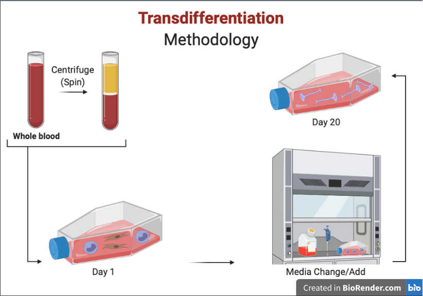
Figure 1. A simplified depiction of Bellon et al.’s4 MDNC methodology. To create MDNCs, researchers first must acquire patient blood. Next, the blood is separated to obtain the peripheral blood mononuclear cells (PBMCs). The monocytes are then separated by centrifugation and magnetism and plated on fibronectin-coated flasks that keep the cells stationary. Over the following days, various chemical compounds are added and removed to help the cells differentiate and survive. Monocytes begin to resemble neurons on day 20.
By day 20, some cells resembled neurons by exhibiting a rounded cell body and long projections that resembled dendrites. Once the monocytes differentiated into neuron-like cells, Bellon et al.4 experimented with different treatments to help identify potential neuronal properties (Figure 2). This is important considering that morphology does not provide much clinical utility other than resembling a neuron. However, if these MDNCs act like neurons, researchers can begin to understand their utility as possible treatments for brain-related diseases. For example, if scientists were to replace damaged neurons with MDNCs in neurodegenerative diseases (i.e., Alzheimer’s disease), MDNCs could function similarly to the original neurons in order to regain brain function.
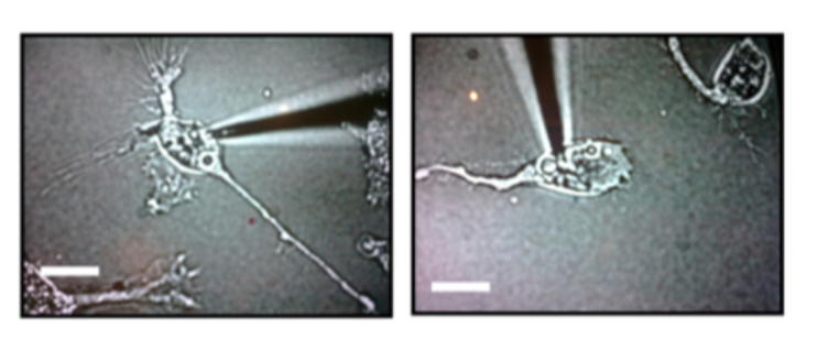
Figure 2. Images from Bellon et al.4. These images were captured using light microscopy. Two MDNCs with rounded cell bodies and dendritic-like projections are pictured above; they were tested for electrical activity with electrophysiology techniques.
Additionally, the differentiated cells contained markers present in normal neurons, suggesting an ability to perform neuron-like tasks. With electrophysiology, Bellon also found that these cells produced electrical signaling, as typical neurons do. So not only do these neuron-like cells look like neurons, but they also act like neurons. Further investigation is needed to identify their full neuronal potential.
Mishra et al.3 were able to take Bellon et al.’s4 study a step further by providing significant findings to this body of research. The retina in our eyes is responsible for receiving and converting light into electrical signals to help us perceive the visual world. A disrupted retina typically leads to blindness, as is the case with retinitis pigmentosa, due to high levels of cyclic guanosine-mono-phosphate (cGMP). Current therapies for this disease only alleviate the symptoms, but cell therapies can regenerate the damaged cells to help them function properly. Mishra et al.3 created retinal neuron-like cells from monocytes with a procedure similar to the one Bellon et al.4 used to create their neurons. Once their cells resembled retinal cells, they transplanted them into the retina of the rodents suffering from retinitis pigmentosa5. The transplanted cells attached to the damaged retinal cells and survived long enough for the rodents to experience improvements in their vision3. After the transplant, there were molecular changes, like decreased cGMP levels, indicating a slight reversal of disease3. Unfortunately, there were also increases in disease-fighting cells (macrophage-related markers) after the transplantation, which can eventually cause the body to fight and remove the transplanted cells over time. It is unclear if this technique will have long-standing positive effects in clinical patients with retinitis pigmentosa; however, these data seem to be a step in the right direction to understand better the disease and how to combat it.
Much work is needed to utilize human blood and its cells as a clinical treatment for brain-related diseases. These data are essential first steps for understanding how monocytes can be made into neurons and how these neurons can then be used to treat various brain-related disorders. However, a standard procedure for differentiation has yet to be established, which would need to be the first step in furthering these data. We found two promising procedures with Mishra et al.3 and Bellon et al.4. Their findings foster hope for procedural replication and discoveries to move forward with one standard procedure. Further understanding of the procedure’s clinical utility is also needed, as the capabilities of these neuron-like cells are undetermined. Mishra et al.3 provided some insight on possible utility for these MDNCs by implanting retinal MDNCs into the retina of blind rodents; however, their findings provided short-term relief of symptoms. Long-term evidence is still needed as well as evidence of utility with other types of brain diseases. So, although your next blood panel may not offer direct insight into your brain just yet, the research promises that new and exciting findings will come from this type of work.
TL;DR
- Blood cells may become a new treatment for brain diseases
- Researchers have learned to make blood cells into neurons
- Neuron-like cells help to cure blindness in rodents
References
- Induced pluripotent stem cells (ips). UCLA Eli & Edythe Broad Center of Regenerative Medicine & Stem Cell Research. (n.d.). https://stemcell.ucla.edu/induced-pluripotent-stem-cells.
- Volarevic, V., Markovic, B. S., Gazdic, M., Volarevic, A., Jovicic, N., Arsenijevic, N., et al. (2018). Ethical and safety issues of stem cell-based therapy. Int J Med Sci., 15(1), 36–45.
- Mishra, A., Mohan, K. V., Nagarajan, P., Iyer, S., Kesarwani, A., Nath, M., … Upadhyay, P. (2020). Peripheral blood-derived monocytes show neuronal properties and integration in immune-deficient rd1 mouse model upon phenotypic differentiation and induction with retinal growth factors. Stem Cell Research & Therapy, 11(1). doi:10.1186/s13287-020-01925-
- Bellon, A., Wegener, A., Lescallette, A. R., Valente, M., Yang, S.-K., Gardette, R., … Hosmalin, A. (2018). Transdifferentiation of Human Circulating Monocytes Into Neuronal-Like Cells in 20 Days and Without Reprograming. Frontiers in Molecular Neuroscience, 11. doi:10.3389/fnmol.2018.00323
- Sahaboglu, A., Paquet-Durand, O., Dietter, J., Dengler, K., Bernhard-Kurz, S., Ekström, P. A., … Paquet-Durand, F. (2013). Retinitis pigmentosa: rapid neurodegeneration is governed by slow cell death mechanisms. Cell Death & Disease, 4(2), e488–e488. doi:10.1038/cddis.2013.12
