By Zekiel Factor
Setting the agenda
Sensation is the bridge between the internal and external world. Examining the elements of processing that our senses have in common allows us to understand how the nervous system gives rise to sensory perception, which is a fundamental component of conscious experience. But what does it mean to go from sensation to sensory perception? This is a subject of endless debate in philosophy and neuroscience, but for our purposes, we’ll use the following specific definitions. Sensation refers to how our bodies transform a stimulus into a neural representation, while sensory perception isour awareness of that neural representation (and, in cases such as hallucinations, does not result from a real sensation at all). It is the latter that allows us to perform further cognitive processes with the sensory information we receive. We use multiple overlapping networks of the nervous system for processing different senses, including areas specialized to integrate information from multiple senses at once. This article will provide an overview of a few key features of this processing: beginning microscopically with a focus on sensory receptors, then following pathways through the brainstem and thalamus to the cortex, and finally zooming out further to interactions at a network level. By examining multiple scales across the scope of this process, we can begin to appreciate how the puzzle pieces of sensory perception fit together.
Sensory receptors, our windows to the world
Sensation enters the body via specialized sensory cells, comprised mostly of different kinds of neurons that relay this sensory information to the brain and spinal cord (AKA the central nervous system; CNS) for processing. Humans are traditionally said to have five senses, but as we learn more about the sensory system, this view has further expanded to subdivide the senses into “general” and “special” categories. Special senses are localized to their own dedicated sensory organ: vision (eyes), hearing and balance (ears), smell (nose), and taste (tongue). General senses, on the other hand, rely on receptors spread all throughout the body: pressure, pain, temperature, movement, and vibration.1
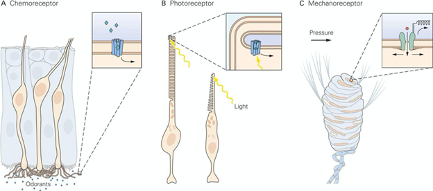
Sensory receptors can be grouped into four functional classes based on what kind of signal they detect: photoreceptors (photons), mechanoreceptors (pressure), thermoreceptors (temperature), and chemoreceptors (chemical ligands).2 All of our senses arise through the transduction of one or more of these stimuli. Some senses utilize multiple subtypes of the same receptor class. Pain, for example, utilizes a combination of different kinds of mechanoreceptors, thermoreceptors, and chemoreceptors. On the other hand, some functional classes of receptors are used for multiple senses, such as mechanoreceptors being used in both hearing (cochlear hair cells in the ear detect frequencies of vibration as sound), as well as in touch (e.g., Merkle cells in the skin detect small amounts of pressure for light touch).
Shared organizational properties of sensory systems
For the most part, senses enter the brain through different regions of the brainstem called “nuclei,” clusters of neurons grouped together by function, with each nucleus tailored to a specific type of sensory information. Touch and body position (trigeminal nuclei), taste (nucleus of the solitary tract), balance (vestibular nuclei), hearing (cochlear nucleus), and some components of vision, all have their initial CNS inputs in the brainstem. In contrast, other components of vision and all of smell (olfactory bulb) have initial CNS inputs elsewhere.4 The nature of these divisions also demonstrate how the unified sensory experience is constructed out of multiple components of each sense, with each component being processed differently. For example, visual information enters the brain in two separate places. The optic nerve sends signals from rod-dominant areas of the eye (more light sensitive) to the upper-most part of the brainstem (superior colliculus of the midbrain), while signals from cone-dominant areas (more color sensitive) bypass the brainstem and go directly to the thalamus (lateral geniculate nucleus).5
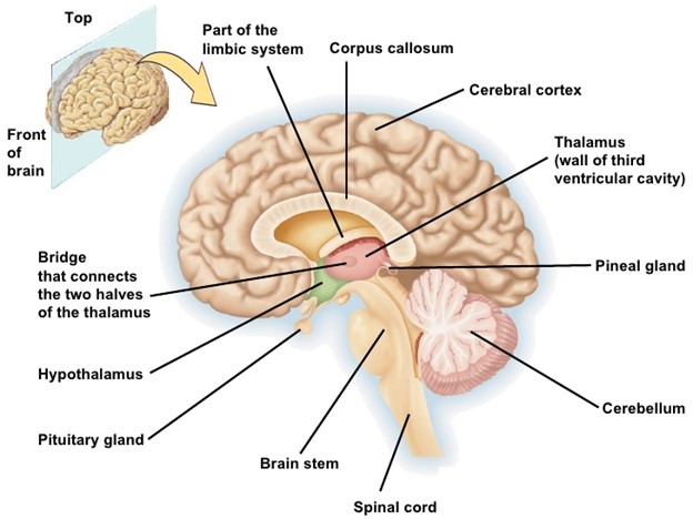
Most sensory information is then routed from the brainstem through the thalamus, which is responsible for relaying most sensory information to the primary sensory areas of the cortex.The thalamus also contains discrete nuclei for processing different senses. 6 It is often described as the sensory processing “gateway” which allows us to filter what sensory information reaches the cortex. The thalamus is constantly being regulated by inputs from other subcortical (“deep brain”) areas like the brainstem and the hypothalamus, as well as the cortex itself. This gating and multiple methods of regulation let the thalamus modulate sensory routing to the cortex in tandem with the sleep-wake cycle and “tune in” more with increased alertness.7
Within the cortex, different areas are specialized to process different sensory inputs: the visual cortex (sight), the somatosensory cortex (touch), the auditory cortex (sound), the gustatory cortex (taste), and the olfactory cortex (smell). Each of these areas contain multiple specialized subdivisions that encode the various characteristics of sensory stimuli. The main sensory processing areas of the cortex share an organized spatial arrangement called topographic mapping. This refers to the way that neurons of sensory pathways, from sense organs all the way to the sensory areas of the cortex, are organized relative to each other. This arrangement localizes the position of a stimulus relative to the body, which gives sensation its spatial component. This spatial component allows us to perceive what direction a sound is coming from, where an injury is located on the body, or where a particular shape is in our field of vision. Importantly, this localization is not always a one-to-one relationship. Input from one sensory receptor may be transmitted to multiple areas of the brain and processed in different ways simultaneously. Disparate inputs from the periphery may share common or overlapping locations in their outputs in the cortex.9 The specifics of how this process takes place in different senses is still an area of major investigation in cognitive neuroscience, and these nuances have critical implications for how each sense is encoded and processed.
How networks piece the world together
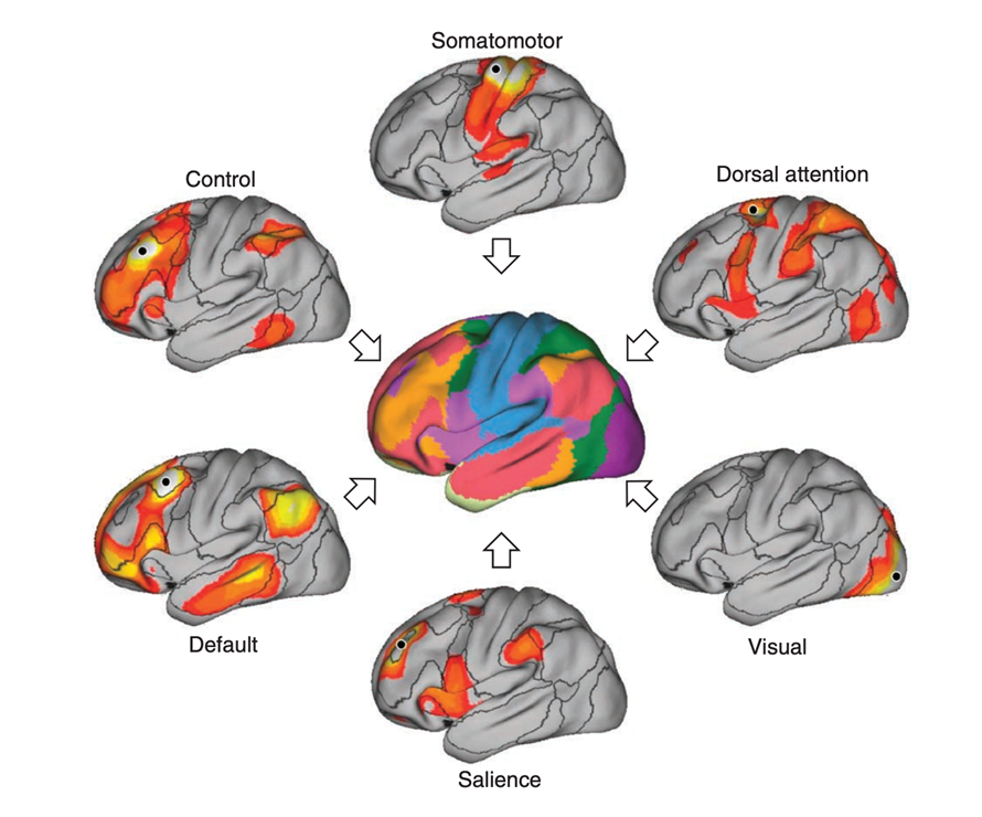
In the field of systems neuroscience, we look at the connections between areas of the brain in terms of networks of activity. Each network is defined by areas which communicate in tandem and exert influence on each other’s activity to perform specific cognitive functions. In general, a network is made up of multiple pathways between the areas of the brain. Neuroscientists have identified many pathways used in processing individual senses and the networks they form. However, our cognition also requires complex cross-talk between multiple networks to refine and transform the information we have taken in for use in specific contexts.11 The default mode network plays a complex modulating role in external focus and alertness and shows high activity during self-referential thinking. The fronto-parietal network is critical for problem solving, and helps us to integrate multiple streams of information to map out the next step to take—both literally in terms of motor planning, and figuratively in terms of goal-directed behavior. The dorsal and ventral attention networks mediate our ability to both direct our attention and to ration it by filtering out the information from the environment we can safely ignore.
Both the thalamus and the sensory processing areas of the cortex maintain important connections with these networks as well as many others and govern the cognitive processing of the sensory information we receive.10 Communication between networks and across senses is what ultimately gives rise to a unified perceptual experience. This encompasses everything from the ability to pay attention to a sudden movement out of the corner of your eye, the ability to assess the environment for danger, or the ability of a song to trigger a memory. The linkage of “bottom-up” (driven by the external environment) processing of sensory information with “top down” cognitive influences (driven by internal mental schemas) is what lets us make sense of, and make judgments about, the information we get from our environment.
In summary, greater than the sum of its parts
Tracing the path of sensory information from the environment through the nervous system offers a window into how this complex array of biological machinery works synergistically, creating a perceptual experience out of the sensations it receives from the world. Sensory receptors gather information by picking up on different kinds of signals (color, temperature, smell) from the environment. Each receptor uses a different mechanism to translate these signals into the common electrochemical language of the brain. Patterns of this electrochemical activity over space and time govern all conscious and unconscious neural processes, forming networks between different areas of the brain that communicate in highly specific patterns to give rise to a coherent sensory experience. Our senses are the most fundamental tools we have for connecting with the world around us, so by understanding their function, we can better appreciate how they change as we develop and age, in different neurological disorders, and the beauty of individual variation in how we perceive the world.
TL’DR:
- Sensation uses many kinds of receptors to detect different kinds of information from the environment.
- Connections between areas of the brain form networks that transform this information into perceptual experience.
Reference
1. Gadhvi, Mahesh. “Physiology, Sensory System.” StatPearls – NCBI Bookshelf, 6 May 2023, http://www.ncbi.nlm.nih.gov/books/NBK547656.
2. Koop, Lindsey K. “Neuroanatomy, Sensory Nerves.” StatPearls – NCBI Bookshelf, 24 July 2023, http://www.ncbi.nlm.nih.gov/books/NBK539846.
3. “Sensory Receptors.” Neuro New Me, 19 Nov. 2020, http://www.neuronewme.com/blog/sensory-receptors-the-basics.
4. Fritzsch, Bernd, et al. “Neurosensory Development of the Four Brainstem-projecting Sensory Systems and Their Integration in the Telencephalon.” Frontiers in Neural Circuits, vol. 16, Sept. 2022, https://doi.org/10.3389/fncir.2022.913480.
5. Gupta, Mohit. “Neuroanatomy, Visual Pathway.” StatPearls – NCBI Bookshelf, 19 Dec. 2022, http://www.ncbi.nlm.nih.gov/books/NBK553189.
6. Torrico, Tyler J. “Neuroanatomy, Thalamus.” StatPearls – NCBI Bookshelf, 24 July 2023, http://www.ncbi.nlm.nih.gov/books/NBK542184.
7. McCormick, David A., and Thierry Bal. “Sensory Gating Mechanisms of the Thalamus.” Current Opinion in Neurobiology, vol. 4, no. 4, Aug. 1994, pp. 550–56. https://doi.org/10.1016/0959-4388(94)90056-6.
8. Islam, Rafiqul. “Thalamus : Anatomy, Location & Function.” Anatomy Info, 3 Sept. 2019, anatomyinfo.com/thalamus-function.
9. Patel, Gaurav H., et al. “Topographic Organization in the Brain: Searching for General Principles.” Trends in Cognitive Sciences, vol. 18, no. 7, July 2014, pp. 351–63. https://doi.org/10.1016/j.tics.2014.03.008.
10. Yuan, Rui, et al. “Functional Topography of the Thalamocortical System in Human.” Brain Structure & Function, vol. 221, no. 4, Apr. 2015, pp. 1971–84. https://doi.org/10.1007/s00429-015-1018-7.
11. Buckner, Randy L., et al. “Opportunities and limitations of intrinsic functional connectivity MRI.” Nature Neuroscience, vol. 16, no. 7, Jun 2013, pp. 832–37. https://doi:10.1038/nn.3423.

This blog has become an invaluable resource for me in navigating the complexities of mental wellness. Thank you for sharing such valuable knowledge!
Itrifal Ustukhuddus