By Mariam Melkumyan
Alcohol use disorder (AUD) is highly prevalent, affecting around 15 million individuals annually in the United States1. The harmful effects of excessive alcohol use on the liver and other organs are well known, however, the exact effect of alcohol on the activity of the different regions and function of the brain is still under study. Numerous conflicting articles exist, with some suggesting that a glass of wine a day is good for you, and others urging that any amount of alcohol can result in immediate negative effects on your brain. But which, if any, of these claims are true?
We still do not know the full effect of occasional alcohol use, as research groups studying the effect of alcohol on various brain regions make new findings every day that raise further questions2–4. My recent publication titled “Astrocytes play a critical role in mediating the effect of acute ethanol on central amygdala glutamatergic transmission” in the Neuropharmacology journal was one of these publications. To understand what the article is about, let’s break down the title and explain each piece separately.
Astrocytes
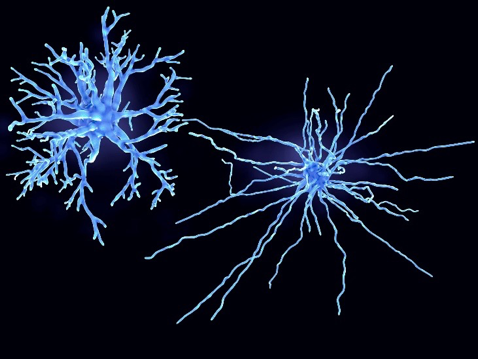
Astrocytes are glial cells and are one of the two main neuroinflammatory cells in the brain5. They are named so, after the Latin word for star, as their projections make them look like stars (Fig. 1).
The main functions of astrocytes include support of neurons through metabolism, transmitter release, maintenance of the blood-brain barrier, and scar formation in case of an injury to the brain tissue5. Astrocytes play a large role in health and disease supporting the integrity of neurons in neurodegenerative diseases and substance use disorders5,6.
Central Amygdala
The central amygdala (CeA) is a brain region responsible for emotional response, fear, and stress. Due to its role in stressful feelings, the circuitry in the CeA has been implicated in withdrawal anxiety of substance/alcohol use and has been shown to be sensitive to drug use2. Alcohol application to the CeA leads to changes in neurotransmission, increasing or decreasing excitability of the cells2,7–9. The experimental changes to the excitability of the cells after alcohol application is dependent on the technique, the concentration of alcohol, and rodent model used, and the subdivision of the CeA being studied, suggesting that this region is highly and differentially sensitive to alcohol2,7. The CeA is divided into two main subdivisions, the medial and the lateral subdivisions, with various cell types in each subregion. My paper focused on the lateral subdivision of the CeA, as this region has not been studied as extensively as the medial subdivision in the context of alcohol use.
Glutamatergic transmission
Glutamate is the primary excitatory neurotransmitter in the central nervous system. Increases in glutamatergic transmission suggest an increase in the excitability of the neurons, meaning that the neurons are more active and transmit more information than at baseline. To study the glutamatergic transmission at a single neuron level, we used whole-cell patch clamp electrophysiology, a technique that allows you to study the electrical properties of neurons at a single neuron level10. When done correctly, whole-cell configuration creates continuity between the cytoplasm and the micropipette (Fig. 2)10, enabling the study of various properties of the cell and its responses to stimuli.

Using electrophysiology, we were able to study the effects of alcohol by recording the spontaneous responses of the cells in CeA brain slices. We saw that when alcohol was applied, there was an increase in the glutamatergic transmission of the cell, suggesting that alcohol increases the excitability of neurons in the lateral subdivision of the CeA. We then asked whether astrocytes play a role in the effect of alcohol on the CeA slices and found that when we blocked astrocytes using a pharmacological blocker and applied alcohol to the slice, there was no change in glutamatergic transmission.
We also asked whether blocking astrocytes chemogenetically would give us the same result as the pharmacological blocker. We used an astrocyte-specific designer receptor exclusively activated by designer drug (DREADDs), which selectively activates the inhibitory metabotropic receptor activity of astrocytes in the presence of a specific drug (here, Clozapine-N-Oxide). This chemogenetic activation of astrocyte inhibitory signaling mimicked the results seen with the pharmacological blocker, confirming that astrocytes mediate the effect of alcohol on glutamatergic transmission in the CeA (Fig. 3).
Putting everything together, the title of the article suggests that when we apply alcohol to a brain slice containing the CeA, we see an increase in the excitability of the neurons in the CeA, and this increase is mediated by astrocyte signaling. Our findings are important as they showcase the involvement of astrocytes in the alcohol-induced increase of glutamatergic activity in the lateral subdivision of the CeA and raise new questions as to whether reducing astrocyte activity can help with AUD. Future studies will need to look at the effect of astrocytes on alcohol dependence to see whether reducing astrocyte activity will lead to reduction in alcohol consumption/seeking. Understanding the role of astrocytes in the effects of alcohol on the CeA and other brain regions can lead to new therapeutics that will allow us to treat AUD.

TL:DR
– Alcohol increases glutamatergic transmission in the lateral central amygdala
– Astrocytes are required for alcohol modulation of glutamatergic transmission
References
1. Understanding Alcohol Use Disorder | National Institute on Alcohol Abuse and Alcoholism (NIAAA). Accessed May 4, 2021. https://www.niaaa.nih.gov/publications/brochures-and-fact-sheets/understanding-alcohol-use-disorder
2. Roberto M, Kirson D, Khom S. The Role of the Central Amygdala in Alcohol Dependence. Cold Spring Harb Perspect Med. Published online January 27, 2020:a039339. doi:10.1101/cshperspect.a039339
3. George F. Koob, Nora D. Volkow. Neurocircuitry of Addiction. Neuropsychopharmacology. Published online 2010. doi:10.1038/npp.2009.110
4. Silberman Y, Winder DG. Ethanol and corticotropin releasing factor receptor modulation of central amygdala neurocircuitry: An update and future directions. Alcohol. 2015;49(3):179-184. doi:10.1016/j.alcohol.2015.01.006
5. Siracusa R, Fusco R, Cuzzocrea S. Astrocytes: Role and Functions in Brain Pathologies. Frontiers in Pharmacology. 2019;10. Accessed January 21, 2022. https://www.frontiersin.org/article/10.3389/fphar.2019.01114
6. Miguel-Hidalgo JJ. The Role of Glial Cells in Drug Abuse. Curr Drug Abuse Rev. 2009;2(1):76-82.
7. Melkumyan M, Snyder AE, Bingaman SS, Arnold AC, Silberman Y. Astrocytes play a critical role in mediating the effect of acute ethanol on central amygdala glutamatergic transmission. Neuropharmacology. Published online December 9, 2021:108918. doi:10.1016/j.neuropharm.2021.108918
8. Silberman Y, Fetterly TL, Awad EK, Milano EJ, Usdin TB, Winder DG. Ethanol produces corticotropin releasing factor receptor-dependent enhancement of spontaneous glutamatergic transmission in the mouse central amygdala. Alcohol Clin Exp Res. 2015;39(11):2154-2162. doi:10.1111/acer.12881
9. Bajo M, Cruz MT, Siggins GR, Messing R, Roberto M. Protein kinase C epsilon mediation of CRF- and ethanol-induced GABA release in central amygdala. Proc Natl Acad Sci U S A. 2008;105(24):8410-8415. doi:10.1073/pnas.0802302105
10. Segev A, Garcia-Oscos F, Kourrich S. Whole-cell Patch-clamp Recordings in Brain Slices. J Vis Exp. 2016;(112):54024. doi:10.3791/54024

Pingback: Get on the write track for an LTS blog post | Lions Talk Science