By Elizabeth Lesko
The history of medical science is long and strenuous, full of great leaps followed by dark ages, brilliant minds tempered by the mores of their time. While we would all like to believe that we live in an enlightened time, full of people who trust the best practices of medical professionals, the current state of affairs in the time of SARS-CoV-19 has made it abundantly clear that the advancement of medical practice is still deeply tied to public opinion. While scientists would like to believe in the power of data to induce positive change, it would prove valuable for us all to consider the power of influence. Art can appeal to a more instinctual, emotional area of the human psyche and great artists are practiced in using their chosen medium to influence their audience towards new ideas. This article will delve into the interplay of art and science through a discussion of the history of human dissection in the Western world as told through visual art.
Classical Greece
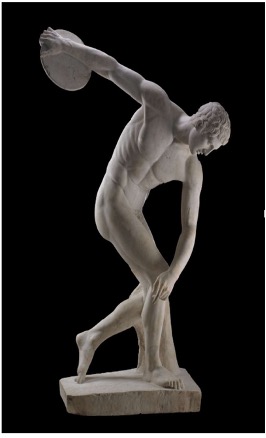
Some of the first recorded studies of the human form date to the Classical era in Greece, where medicine was intertwined with philosophy. During this era dissection was performed primarily to answer questions that were more religious than medical in nature. Around the 4th century BC, the Greek philosopher Hippocrates is widely credited with the birth of medicine as a discipline distinct from philosophy1. The Roman physician Galen further developed Hippocratic medicine in the 2nd century AD. At this time the dissection of human bodies was considered taboo by the Greeks and Romans, but a strong interest in human anatomy persisted. Physician-philosophers such as Galen were known to follow in the tradition of Aristotle’s studies on biology and dissect animals such as monkeys and pigs2. The information gained from such dissections was supplemented by study of the superficial anatomy of human corpses when available. This era’s fascination with the human form is reflected in the detailed yet sometimes inaccurate anatomy of Greek sculptures (Figure 1)3, suggesting that artists likely studied anatomy using the same resources available to physicians during this foundational age of medicine.
Middle Ages

Following the establishment of Christianity and the Holy Roman Empire in 10th century AD, medicine as a distinct and scientific discipline began to stagnate. Religious morality of the time balked at the idea of desecrating the human body by dissection and much medical knowledge accrued during the first centuries AD was forgotten or ignored4. Medicine remained a distinct discipline practiced by physicians, but most experiments were only repetitions of those performed by Galen or Aristotle. Most anatomical knowledge was based on examination of injuries (Figure 3) and diseases with external symptoms until the establishment of the first universities in the 12th century.

The demands of universities for new information on human anatomy to aid the development of medicine and surgery was answered by decrees from the Pope – the highest European authority on morality at the time – allowing for some rare human dissections exclusively for training purposes4. These dissections would be few and far-between and, consequently, well-attended by physicians and the public alike. Western paintings and sculptures during the Middle ages was almost exclusively religious and stylized, though a few images depicting studies of human anatomy can be found (Figure 2).
Renaissance and Early Modern Era

The Renaissance continued in the tradition of public dissections established during the late Middle Ages, with many artists attending public dissections or even performing their own to better understand the human form. Leonardo da Vinci and Michelangelo famously performed many dissections during their respective anatomical studies and made detailed observations and drawings (Figures 4 and 5)4. In many parts of Europe human dissection was still under strict limitations, allowing only the dissection of convicted criminals and limiting the number that could be legally performed by a given institution.

Physicians of this period were also known to generate their own works depicting human anatomy with the assistance of a skilled artist, such as the famous De humani corporis fabrica by Andreas Vesalius (Figure 6)5. This book is considered both a continuation and discreditation of Galen’s work on anatomy and contains many detailed and strikingly artistic renderings of the human form in various states of dissection.

While public dissections emerged as a mainstay of anatomical teaching during the Renaissance, the rarity of such an opportunity made these anatomy lessons into macabre social events often attended by the wealthy in addition to medical students4. The underprivileged were unlikely to ever have the opportunity to attend such an event themselves and, perhaps in consequence, were more likely to consider the practice horrific and immoral. Depictions of dissections in artworks of the time were rare but may have had an impact in generating broader acceptance of the practice. A striking example of this is Rembrandt’s The Anatomy Lesson of Dr. Nicholaes Tulp, a fictitious rendering of a real dissection attended by the artist (Figure 7)6.
18th, 19th, and Early 20th Century
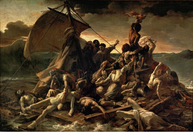
While acceptance of human dissection had been firmly established in the medical world by the 18th century, much of the public still considered dissection disrespectful to the individual. Cadavers were therefore sourced strictly from executed criminals (as an additional punishment) or, later in the 18th century, from the unclaimed dead4. Rising demand for cadavers from rapidly growing medical schools across the Western world meant that these sources eventually proved insufficient and grave robbing of freshly buried individuals by “resurrection men” for profit became increasingly problematic. The Anatomy Act of 1832 in England officially legalized the use of unclaimed bodies of the poor from workhouses and hospitals while simultaneously forbidding the use of executed individuals and allowing voluntary donations. Art during this period was rapidly developing and expanding in subjects beyond depictions of the human form, but artists such as Théodore Géricault were still known to study cadavers or disembodied parts in the pursuit of realism (Figure 8)7.

The 19th century continued the development of medical texts containing detailed renditions of human anatomy with the publication of Gray’s Anatomy in 1858 (Figure 9)8. This momentous text was a direct consequence of the Anatomy Act, as cadavers were now more easily obtained for study. While valuable to the pursuit of science and medicine, the limitations on subjects for dissection led to unethical practices in obtaining bodies4. The poor were less likely to be able to afford burial or take action against body-snatching, and the bodies of slaves were sold to teaching physicians without thought to the desires of the deceased person’s family. Psychiatric patients who died in asylums also disproportionally ended up on a dissection table.
Late 20th Century and 21st Century

Questionable sourcing of subjects for human dissection peaked in the mid-20th century in the midst of the atrocities of World War II, forcing a drastic change in policy post-war. Legislation was established to allow the voluntary donation of bodies for anatomical teaching and was refined over the latter half of the 20th century4. Meanwhile, great advancements in other areas of science and medicine improved public opinion of medical research and allowed body donations to meet the demand for cadavers in medical training. Gone are the days of public dissections – anatomical teaching is now firmly the realm of private schooling for the purpose of training physicians. Human dissection still has its place in modern art, however. It is common for artists to study anatomy textbooks and live figures for an understanding of the human figure, and some individuals such as artist Damien Hirst even seek to directly use anatomy and dissection as art to inform or influence their audience (Figure 10).
A more infamous example of dissection in modern art is the “Real Bodies” travelling exhibit that used plasticized cadavers in lifelike poses to display the human form. This exhibit also demonstrates that the controversies of human dissection are not in the past, as it is believed the cadavers used in the exhibit may have been executed prisoners originating from China9.
Reading accounts of old-fashioned morality impeding scientific progress can be frustrating as a scientist, but the “Real Bodies” controversy serves as a poignant reminder that ethical considerations are still incredibly important in protecting the rights of all people – living or dead. The preeminent force driving someone’s opinion on – for instance – the Covid-19 pandemic responses can depend just as heavily on their morals as on facts or data. Considering an audience’s values is an absolutely vital skill for a scientist to communicate their work to the public, and I believe that studying how artists have utilized and expressed such revolutionary ideas as human dissection is an invaluable way to build such a skill.
References
- FH Garrison. History of Medicine. Philadelphia: W.B. Saunders Company. 1966.
- AJ Brock (translator). Introduction. Galen. On the Natural Faculties. Edinburgh. 1916.
- FN Pryce, AH Smith. Sculpture / Catalogue of Greek Sculpture in the British Museum. BMP, London. 1892-1928. https://www.britishmuseum.org/collection/object/G_1805-0703-43
- SK Ghosh. Human cadaveric dissection: a historical account from ancient Greece to the modern era. Anat Cell Biol. Sep 2015. 48(3): 153–169.
- CD O’Malley. Andreas Vesalius of Brussels, 1514-1564. Berkeley: University of California Press. 1964.
- H Rachlin. Scandals, Vandals and Da Vincis. Chrysalis Books. 2007. 55–61.
- R Christiansen. The Victorian Visitors: Culture Shock in Nineteenth-Century Britain. New York Times. 3 June 2001.
- H Gray, HV Carter. Anatomy Descriptive and Surgical. London: John W. Parker and Son. 1858.
- F Mao. ‘Real bodies’ exhibition causes controversy in Australia. BBC News. 26 April 2018.
Figure 1: The British Museum (https://www.britishmuseum.org/collection/object/G_1805-0703-43)
Figures 2-10: Wikimedia commons

Pingback: 03 – Pre-Production – Paige Jenkins – Major Project