By Rebecca Fleeman
Technology has significantly changed the way we interact with our environment. One noticeable advance is technology continuously becoming smaller and smaller over time. For example, look at how we listen to music. We began with records, moved to cassette tapes, then ended on CDs before everything we listened to became stream-able on the internet. Likewise, computers have gone from taking up whole rooms to fitting in each student’s portable backpack. Biomedical research has followed suit in the quest to minimize materials needed to study human health. Typically, research is conducted from bench to bedside in a methodical matter: in vitro studies of biochemistry, moving to cell lines, then creating an in vivo animal model, testing in a secondary mouse model, then moving through to clinical trials. This process is lengthy, and can take upwards of ten years, not to mention millions of dollars1. But as in the past, technology has again given way to a smaller, more practical method: cue organ on a chip.
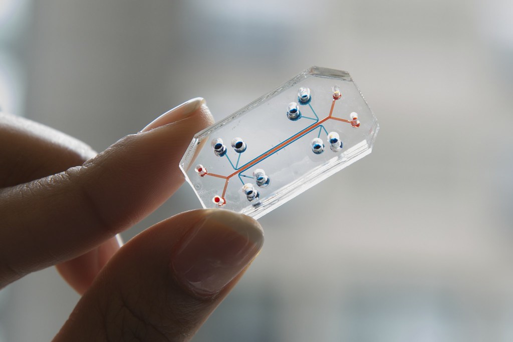
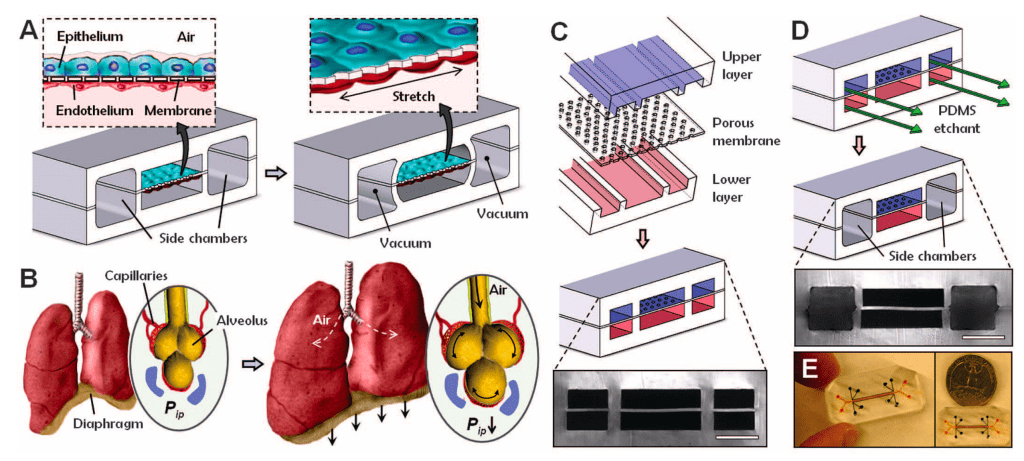
The concept of organ on a chip began in the 1990s when Manz et al. created the first miniaturized total chemical analysis system2. This ultimately paved the way for the field of microfluidics, which is the study of geometrically constrained fluid systems in a microscale environment. Scientists take advantage of this microscopic system to scale down experiments into USB-drive-sized chips that resemble computer microchips3. The first organ on a chip creation was credited to the team of Dr. Donald Ingber and was published in Science in 20104. Ingber is the founding director of the Wyss Institute at Harvard, specializing in bioengineering. Since his first organ on a chip, the lung, research teams have gone on to create organ on a chip systems for the skin, kidney, intestine, placenta, brain, and more3. While these highly advanced tools do not recapitulate the entire organ, they can be used to accurately test a myriad of organ functions and their responses to drugs and environmental factors.
So how do they work? First, the microfluidic channel chip is made (this is now often carried out through 3D printing techniques). Next, the chip is inoculated with cells. This step allows for the large amount of customization in the model, with a number of cell types have been used, such as epithelial, endothelial, smooth muscle, and organ-specific cells2. In fact, multiple cell types can be added to allow for a more systemic environment. Once cells have been placed in the chip, grown, and differentiated, they will develop their full cell function potential to operate as the desired organ. From here, the chip which resembles key functions of the organ of choice can be challenged with different therapeutics, toxins, or biologic compounds to measure responses.

What is the future of organ chips, and will organ chips ever fully replace pre-clinical animal models of studying drug development and disease? The Wyss Institute has made some extremely impressive advances that point towards a greater amount of organ chips in the future of drug development. Recently, this team even figured out how to link multiple organ chips through vascular systems, which mimic a whole-body chip. Organ chip models have been made for almost every organ and utilized to study a countless diseases such as multiple cancers, Alzheimer’s disease, cardiovascular disease, and viral infections2. Now that the organ chip technology is more widely available, the next steps are creating a greater number of functions that can be tested in these devices so that we can shorten the time for drug development and discovery5. Unfortunately, the technology is still expensive so deciphering ways to minimize costs will also be important to the future of organ chips5. While many hope that these chips will entirely replace animal testing, we are still quite a ways away from this dream. Because these chips cannot completely replicate the complexities of a multifunctional organ, in vivo studies are still required for rigorous testing5. However, as this technology develops, you can imagine how many years, dollars, and animal lives we could potentially save by implementing organ chips more into the field of biotechnology.
1. Dickson, M. & Gagnon, J. P. Key factors in the rising cost of new drug discovery and development. Nat. Rev. Drug Discov. 3, 417–429 (2004).
2. Sosa-Hernández, J. E. et al. Organs-on-a-Chip Module: A Review from the Development and Applications Perspective. Micromachines 9, (2018).
3. Ingber, D. E. Developmentally inspired human ‘organs on chips’. Development 145, (2018).
4. Huh, D. et al. Reconstituting Organ-Level Lung Functions on a Chip. Science (80-. ). 328, 1662–1668 (2010).
5. Wu, Q. et al. Organ-on-a-chip: recent breakthroughs and future prospects. Biomed. Eng. Online 19, (2020).
