By Sarah Latario
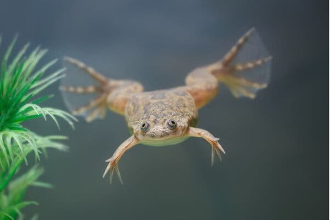
For over three decades, African clawed frogs were the embodiment of pregnancy tests, rather than the small kits found in the drug store. After the British scientist Lancelot Hogben discovered that injecting urine samples from pregnant women caused the frogs to lay eggs, they became the standard for pregnancy tests across the globe from the 1930s to the early 1960s.1 Hogben’s discovery occurred after he moved to South Africa and began using the local African clawed frogs, part of the Xenopus genus, for his research on hormones. These frogs were abundant and easy to work with, allowing him to smoothly continue his work on endocrinology. During one of his experiments, he injected ox pituitary extract into one of these frogs, unexpectedly causing it to lay eggs. It was already known that the urine from pregnant women contained pituitary hormones, sparking Hogben to realize these frogs could be used as pregnancy tests. Shortly after this discovery, he returned to England, bringing the frogs with him to further tease out this idea. After verification experiments, Hogben determined that if urine samples from pregnant women were injected into the frogs it caused them to lay eggs within 5 to 18 hours post-injection. During this preliminary research, one of the lab technicians conducted 2,112 tests and did not have “one clear positive that did not indicate a pregnancy. There were a few negative results which when repeated after a fortnight became positive, but I do not think that these can be regarded as failures”. This technique was considered by physicians to be the most dependable pregnancy test for decades and was only ended when immunological testing was discovered in the 1960s. Current pregnancy tests and the “Hogben test” both work by the same mechanism, recognizing human chorionic gonadotrophin (hCG) in the urine of pregnant women. Although no longer used in pregnancy testing, this phenomenon laid the groundwork for the African clawed frog (Figure 1) being widely available as a model organism for future researchers.
Since Hogben brought them out of South Africa, Xenopus have been used globally in research labs and have been involved in 21 of the last 91 Nobel-Prize-winning research studies that used animal models. It is estimated that about 90% of human-disease-associated genes have Xenopus homologs, making them closer to humans than zebrafish and C. elegans. 6, 9, 10 Additionally, African clawed frogs are sensitive to many hormones, aside from hCG, a quality that has greatly increased their use by, and value to, researchers across disciplines. For example, the frogs have been used extensively to study thyroid hormones (TH), as their transition from a tadpole to a mature frog is completely dependent on the activity of TH.2,3 Xenopus have also been used in sex hormone studies to identify the reproductive toxicity of endocrine disruptors.4 The discovery of the hormone sensitivity of these frogs has opened the door for researchers at numerous institutions, as well as the United States Environmental Protection Agency, to utilize them in environmental toxicology studies.3 This hormone sensitivity makes Xenopus a strong model organism to study not only the role of hormones in development, but also the presence of natural and artificial hormones in the environment and the effects these may have on wildlife and humans. Environmental estrogens, hormones that do not naturally come from the human body and range from synthetical chemicals to natural plant compounds, are a type of endocrine disruptor that is often studied using Xenopus models.5 Additionally, thyroid disruptors – one of the most prevalent environmental estrogens– have also been shown to disrupt Xenopus early development.3
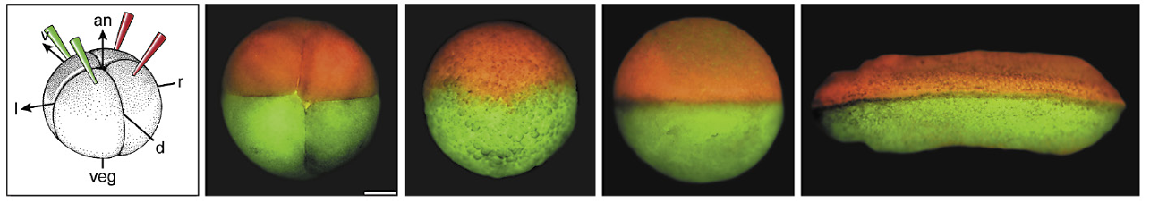
Beyond their role as early pregnancy indicators and use in hormone studies, African clawed frogs have also played crucial roles in research in the field of development and physiology. Development stages occurring during the first 1-2 days of frog embryonic growth would take weeks to achieve in a mouse model. The further value of using Xenopus in developmental studies can be attributed to their embryos being large and easy to microinject. The embryos also remain consistent in size as early development progresses, preventing the dilution of experimental substances as the frog increases in mass. During experiments, nucleic acids, proteins, and whole nuclei and organelles can be easily injected into either the whole frog embryo or specific cells. Cell-free extractions can also be easily removed from immature egg cells to perform in vitro biochemistry and molecular biology. After the first cellular cleavage, the midline of the frog forms where the body is divided in half and separates, allowing for one side to be injected with a mutated gene or pharmaceutical while leaving the other side unaltered (Figure 2).6 With these one-sided injections, a single frog can have both the control and experimental in the same organism, decreasing variability.6 Furthermore, Xenopus embryos can tolerate dramatic manipulations such as transplantation of single cells and ‘cut and paste’ of different larger sections of the embryo.3 ‘Windows’ have even been made within living embryos where the outer section of the embryo is removed to allow for live imaging of the kidneys as they develop over time.7
Some of the additional research areas in which Xenopus are most utilized are physiological studies of the musculoskeletal, cardiovascular, renal, respiratory, reproductive, and sensory systems as well as evolutionary biology.8 The African clawed frog model organism has also been used as a tool to study cellular processes like cell movement, cell fate, cell cycle, the cytoskeleton, chromatin, ion channels, and apoptosis. The Xenopus genus has both diploid (X. tropicalis) and allotetraploid (X. laevis) species, often used in genetic research. These frogs have been widely used to discover many different genes involved in congenital heart defects, kidney disease, cancer, gastrointestinal and pancreatic disorders, neurological diseases, muscle atrophy, and human ciliopathies.3
Overall, Xenopus make a strong vertebrate model to study a myriad of processes and systems across diverse disciplines – in other words, these frogs are more than just frogs. Relied upon for decades as a reliable pregnancy test and vertebrate research model, the enormous contributions made by the African clawed frog towards human disease research should not be forgotten.
TL:DR
- Xenopus frog models have been used for decades in many scientific disciplines.
- Made popular as pregnancy tests, they are still frequently used in current research in diverse fields.
References
1. Pearl, E. et al. An optimized method for cryogenic storage of Xenopus sperm to maximise the effectiveness of research using genetically altered frogs. Theriogenology 92, 149–155 (2017).
2. Damjanovski, S. et al. Role of ECM remodeling in thyroid hormone-dependent apoptosis during anuran metamorphosis. Ann N Y Acad Sci 926, 180–191 (2000).
3. Liu, L. S., Zhao, L. Y., Wang, S. H. & Jiang, J. P. Research proceedings on amphibian model organisms. Zool Res 37, 237 (2016).
4. Qin, Z. & Xu, X. Application of Xenopus laevis in ecotoxicology (I) —Introduction and quality control of laboratory animal. Chinese Science Bulletin 2006 51:11 51, 1273–1280 (2006).
5. Bevan, C. L., Porter, D. M., Prasad, A., Howard, M. J. & Henderson, L. P. Environmental estrogens alter early development in Xenopus laevis. Environ Health Perspect 111, 488 (2003).
6. Blum, M. & Ott, T. Xenopus: An Undervalued Model Organism to Study and Model Human Genetic Disease. Cells Tissues Organs 205, 303–313 (2018).
7. Krneta-Stankic, V. et al. The Wnt/PCP formin Daam1 drives cell-cell adhesion during nephron development. Cell Rep 36, (2021).
8. Burggren, W. W. & Warburton, S. Amphibians as Animal Models for Laboratory Research in Physiology. ILAR J 48, 260–269 (2007).
9. Baldridge, D. et al. Model organisms contribute to diagnosis and discovery in the undiagnosed diseases network: Current state and a future vision. Orphanet J Rare Dis 16, 1–17 (2021).
10. Howe, K. et al. The zebrafish reference genome sequence and its relationship to the human genome. Nature 496, 498 (2013).

Pingback: Discover The Mystical Albino African Clawed Frogs
wow what a informative blog