By: Paige Bond
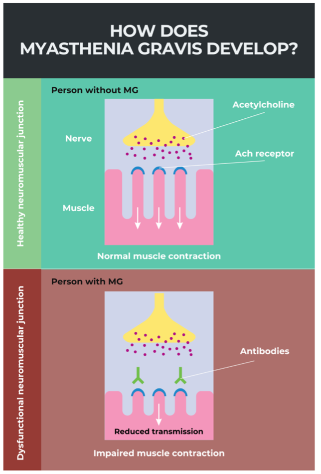
Movement requires communication between both muscles and nerves. At the neuromuscular junction (Figure 1), nerves release a chemical messenger, acetylcholine, which binds to specific receptors on muscle cells to trigger muscle contraction. Disrupting the activity of the acetylcholine receptors can lead to muscle weakness or the inability to voluntarily contract muscles at all. In autoimmune myasthenia gravis (MG), the patient’s immune system prevents acetylcholine from binding to the acetylcholine receptors by damaging the receptors or receptor-associated proteins on muscle cells, leading to impaired muscle contraction. Although MG is a rare and complex disease, estimates from 2023 predict the prevalence of myasthenia gravis to be about 20 out of 100,000 individuals worldwide.
There are multiple subtypes of autoimmune MG. Most individuals first develop ocular MG, leading to drooping eyelids, eye weakness, and double vision. Half of the individuals diagnosed with ocular MG later develop generalized MG which can expand the ocular muscle weakness to the face, neck, arms, legs, and throat. 15-20% of individuals with MG also experience a myasthenic crisis at least once in their lives. A myasthenic crisis results from severe respiratory weakness and requires immediate emergency care. For individuals affected with MG, better therapeutic options than the ones currently available will not only result in better quality of life but can also save their life.
Basics of the immune system
In healthy individuals, the cells within the immune system eliminate foreign pathogens such as bacteria and viruses, as well as damaged or infected cells. There are two arms of the immune system: innate and adaptive. The innate immune system acts quickly. It is the first line of generalized defense against pathogens, causing the familiar symptoms of inflammation and fever. In contrast, the adaptive immune system takes longer to respond. Cells of the adaptive immune system have a much more targeted response toward an antigen, a small piece of a pathogen that can generate an immune response.
In autoimmune disorders, the adaptive immune system reacts to self-antigens1. These molecules, as the name suggests, are from the body and are not foreign invaders; they should not generate an immune response. However, in autoimmune disorders, the adaptive immune system loses self-tolerance (for an explanation of how self-tolerance can be lost, read Niesha Savory’s LTS article “Reprogramming your defenses – autoimmune disease and a ‘small’ potential therapy”), causing the immune cells to target otherwise healthy cells in the body.
During a normal immune response following an infection, B and T cells, the cells of the adaptive immune system, mount a response against invading pathogens. Plasma cells—mature, differentiated B cells—secrete specialized proteins called antibodies that bind to the surface of pathogens. Antibodies binding to pathogens can both prevent their entry into host cells and induce their phagocytosis (engulfment) by innate immune cells such as macrophages. While B cells prevent infection of new cells and can directly limit infection spread, cytotoxic CD8 T cells secrete molecules which directly kill infected or damaged cells. In autoimmunity, plasma cells secrete antibodies targeted at self-antigens called autoantibodies2, whereas cytotoxic CD8 T cells target normal tissue. These two arms of the adaptive immune system show that when cells of the immune system are wrongly targeted towards the host, it creates a cycle of damage and inflammation that leads to autoimmune disorders. In the case of autoimmune MG, the immune system generates autoantibodies that recognize and bind to the acetylcholine receptor and associated proteins at the neuromuscular junction (Figure1). Binding of the autoantibodies to these proteins contributes both to blocking and destroying the acetylcholine receptors, inhibiting communication between nerves and skeletal muscles.
Current therapies for myasthenia gravis
The first line of therapy for MG involves increasing the amount of acetylcholine present at the neuromuscular junction, often with acetylcholinesterase inhibitors such as pyridostigmine3. The inhibitor works by stopping acetylcholinesterase, the protein responsible for degrading acetylcholine, resulting in more acetylcholine at the neuromuscular junction. Other therapies aim to reduce the IgG autoantibody levels in MG patients by inhibiting the neonatal Fc receptor (FcRn)4. FcRn is a specialized protein that stabilizes antibodies like IgG, which supports their role in long-term immunity. IgG antibodies are the most specific type of antibody made and are found at the highest concentration in the body. They are important for the secondary immune response, or fast adaptive response during reinfection or whenever the body encounters a familiar antigen. A major limitation of FcRn inhibitors is that MG patients require treatment every two weeks, putting strain on patients that do not have easy access to medical care5. Additionally, many patients undergoing FcRn inhibitor administration still have MG symptoms, which is why most FcRn treatments also supplement steroid medication6. Unfortunately, long-term steroid use causes broad immunosuppression and can make MG patients more susceptible to other diseases as well as reduce the efficacy of vaccines7. Therefore, there is a need for new therapies that have more long-term efficacy and do not have broad immunosuppressive effects. The Descartes-08 trial, named in the Nature Medicine Year in Review: Eleven clinical trials that will shape medicine in 2026, is a CAR T cell therapy that aims to do just that while alleviating MG symptoms.
The basics of CAR T cell therapy
Chimeric antigen receptor (CAR) T cell therapy is an individualized treatment that utilizes a patient’s T cells and is mostly used to treat different types of cancer. To generate CAR T cells, T cells are isolated from the patient and genetically manipulated to express a synthetic cell surface receptor. The T cells use the synthetic receptor to target and deplete a specific cell type. For multiple myeloma, a cancer of plasma cells, CAR T cell therapies are used to target the B cell maturation antigen, or BCMA8. BCMA is a protein expressed on the cell surface of long-lived plasma cells and is required for their survival; it is expressed in normal and malignant myeloma plasma cells9. To treat this cancer, CAR T cells were engineered to express anti-BCMA which directs CAR T cells to deplete the target plasma cells by binding to BCMA. Some consequences of traditional anti-BCMA CAR T cell therapy is that it causes a decrease in patients’ healthy plasma cell population, requires lymphodepletion (a chemotherapy that depletes the patient of native immune cells), cannot be administered in an outpatient setting, and can be toxic. The most common example for toxicity in CAR T cell therapy is cytokine release syndrome, during which effector immune cells become overactive, resulting in systemic inflammation10. Researchers behind the Descartes-08 CAR T cells aim to circumvent these limitations by using mRNA instead of genetically manipulating the CAR T cell DNA.
Descartes-08, a novel mRNA CAR T cell therapy
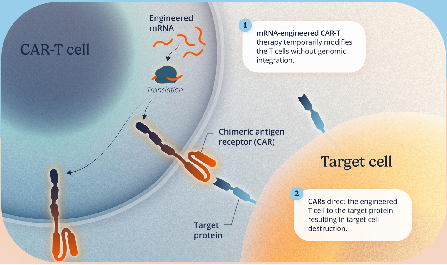
Descartes-08 CAR T cells also target BCMA; however, the novel therapy starts with mRNA, the immediate downstream molecule of DNA that gets transcribed before being translated into a functional protein (Figure 2). Compared to traditionally engineered CAR T cells that have the patients’ T cell DNA permanently genetically modified, Descartes-08 utilizes mRNA which is added into the patient’s T cells. Use of mRNA does not result in permanent modification of the genetic code of the patient’s T cells. The product of anti-BCMA mRNA CAR T cells is the same as traditionally engineered CAR T cells because the downstream biological process between both Descartes-08 and traditionally engineered CAR T cells to generate the synthetic receptor is the same. Due to the use of mRNA administration, Descartes-08 CAR T cells have added benefits to address challenges associated with CAR T cell therapies. Most importantly, adding the synthetic receptor at the mRNA level adds a self-regulating proliferation limit to the Descartes-08 CAR T cells. As the cells divide, the mRNA decreases overtime. This contrasts with traditionally engineered CAR T cells, where the integrated DNA would be passed down with each cell division. Major benefits of the transient targeting of BCMA in the Descartes-08 CAR T cells are that they can be administered without lymphodepletion and in an outpatient setting, making the therapy more accessible. These benefits support Descartes-08 as a safer alternative to previous BCMA-directed therapies that have toxicity issues11.
The Descartes-08 Study Setup
Before becoming available to the public, therapies must make it through three phases of clinical trials. Descartes-08 just completed its phase 2b trial, so it is still in the middle stages of approval. The study consisted of 26 MG patients: 15 received the Descartes-08 and 11 received placebo in a double-blind, randomized trial6. Patients received a weekly infusion of either CAR T cells or placebo for six weeks total. One important limitation of the Descartes-08 study to note is that many of the participants were on moderate levels of steroids and had received other MG therapies before the trial6.
The Descartes-08 Study Results
To test the efficacy of the CAR T cell therapy, researchers looked at different molecular and clinical readouts5,6. For immune therapies, characterizing different immune cell populations as well as their activity identifies the basis of what is happening in the patients at a cellular level. MG patients have a different baseline of immune cell activation compared to healthy individuals, with increased activation of two immune cell types linked to autoimmunity: early-stage plasma cells (PCs) and plasmacytoid dendritic cells (pDCs)5. Elevated activity of these cells signals ongoing immune function. Indications of Descartes-08 administration being successful at calming an overactive immune system would be observing decreased immune cell activation and seeing heightened BCMA expression returning to healthy low levels found in non-MG patients. Months after the trial, patients that received Descartes-08 CAR T cells had decreased activation of PCs and pDCs, while the overall number of target cells which give rise to PCs and pDCs did not change5. Importantly, these results indicate that Descartes-08 was specifically able to reduce BCMA levels without significantly dampening the overall immune response.
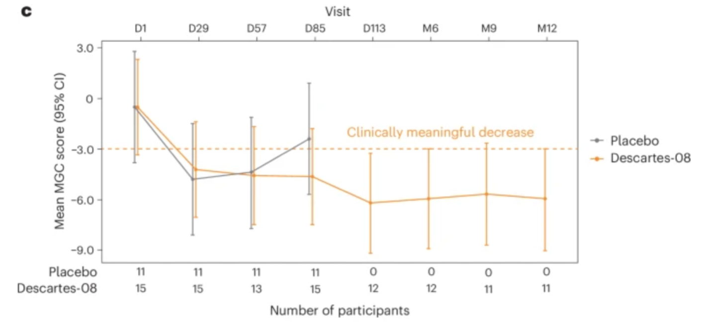
To investigate clinical outcomes, patients were evaluated using the MG Composite (MGC) score6, a clinical point scale that physicians use to evaluate symptoms of individuals with MG. For phase 2b of the Descartes-08 trial, the primary goal was to have the MGC score improve by decreasing greater than or equal to five points by the third month of the clinical trial. Researchers noted a significant decrease in the MGC score for patients that had been administered Descartes-08 compared to placebo, an improvement that persisted months after the trial was completed (Figure 3)6. Patients that took Descartes-08 were also allowed to reduce prednisone usage after month six. The median patient steroid dose at month 12 of the trial was 55% lower compared to their dosage at the beginning of the study6. There were a high number of minor adverse events noted such as acute headache, nausea, and low fever, but most of these events resolved within 24 hours and did not require hospitalization. Overall, these data show that patients administered Descartes-08 showed a significant improvement in MG symptoms with only minor side effects compared to those administered the placebo.
So, what’s the big deal?
Although MG is considered a rare disease, there are more than one million individuals around the world living with MG. The prevalence for the number of individuals living with MG is estimated to be around 150 to 200 per every million individuals in 202112. Descartes-08 would improve symptoms of MG without resulting in broad overall immune suppression, allowing these individuals to live more normal lives. The availability of Descartes-08 in an outpatient setting makes treatment more accessible, a major shortcoming of many CAR T cell therapies currently available6. Importantly, taking what they have learned in the Descartes-08 trial, researchers are expanding to treat other autoimmune diseases such as myositis and systemic lupus erythematosus. These data suggest that mRNA therapies are not only useful for effective vaccine development; they may be useful in expanding CAR T cell therapy beyond cancer treatments to help individuals with autoimmune diseases.
TL; DR:
- Myasthenia gravis (MG) is an autoimmune disorder that disrupts communication between nerves and muscles, which can be life-threatening.
- Descartes-08 is a novel mRNA CAR T cell therapeutic that may be safer and more accessible for individuals with MG than traditional CAR T cell therapies.
References
- Yasmeen, F.; Pirzada, R. H.; Ahmad, B.; Choi, B.; Choi, S. Understanding Autoimmunity: Mechanisms, Predisposing Factors, and Cytokine Therapies. Int. J. Mol. Sci. 2024, 25 (14), 7666. https://doi.org/10.3390/ijms25147666.
- Malkiel, S.; Barlev, A. N.; Atisha-Fregoso, Y.; Suurmond, J.; Diamond, B. Plasma Cell Differentiation Pathways in Systemic Lupus Erythematosus. Front. Immunol. 2018, 9, 427. https://doi.org/10.3389/fimmu.2018.00427.
- Gilhus, N. E.; Andersen, H.; Andersen, L. K.; Boldingh, M.; Laakso, S.; Leopoldsdottir, M. O.; Madsen, S.; Piehl, F.; Popperud, T. H.; Punga, A. R.; Schirakow, L.; Vissing, J. Generalized Myasthenia Gravis with Acetylcholine Receptor Antibodies: A Guidance for Treatment. Eur. J. Neurol. 2024, 31 (5), e16229. https://doi.org/10.1111/ene.16229.
- Ulrichts, P.; Guglietta, A.; Dreier, T.; Van Bragt, T.; Hanssens, V.; Hofman, E.; Vankerckhoven, B.; Verheesen, P.; Ongenae, N.; Lykhopiy, V.; Enriquez, F. J.; Cho, J.; Ober, R. J.; Ward, E. S.; De Haard, H.; Leupin, N. Neonatal Fc Receptor Antagonist Efgartigimod Safely and Sustainably Reduces IgGs in Humans. J. Clin. Invest. 2018, 128 (10), 4372–4386. https://doi.org/10.1172/JCI97911.
- Fedak, R. R.; Ruggerie, R. N.; Shan, Y.; Curvino, E. J.; De Sousa, J. F.; Daniel, S.; Ngo-Casi, M.; Kamboh, H.; Vu, T.; Durmuş, H.; Mozaffar, T.; Howard, J. F.; English, E. P.; Benson, A.; Duvernay, M. T.; Singer, M. S.; Kalayoglu, M. V.; Brunn, C.; Bodansky, A.; Anderson, M. S.; DeRisi, J. L.; Garcia, S. T.; Yu, D. J. L.; Zorn, K. C.; Kurtoglu, M.; Miljković, M. D.; Stewart, C. A.; Jewell, C. M.; on behalf of the MG-001 Study Team; De Sousa, J. F.; Buensuceso, C.; Tostanoski, L. H.; Zhang, Y.; Xu, H.; Pope, C. A.; Schneider, D.; English, A.; Filev, A.; Biaksangi, J.; Small, G.; Rivner, M.; Badruddoja, M. A.; Myers, B.; Shroff, S.; Slanksy, A.; Holley, K.; Sahagian, G.; Quesnell, T.; Feinberg, M. H.; DeMaria, G.; Vullaganti, M.; Barnett-Tapia, C.; Mendoza, M.; Irannejad, M.; Siddiqi, Z.; Habib, A. A.; Ragole, T.; Pasnoor, M.; Traub, R. E.; Chopra, M.; Grover, N. S.; Park, Y. A.; Karafin, M.; Trunnell, A.; Cheng, C.; Karam, C.; Davis, D.; Shaw, J.; Requena, N.; Peltier, A.; Gwathmey, K. BCMA-Directed mRNA CAR-T Cell Therapy for Myasthenia Gravis: Exploratory Biomarker Analysis of a Placebo-Controlled Phase 2b Trial. Nat. Med. 2026, 32 (3), 1118–1130. https://doi.org/10.1038/s41591-025-04170-z.
- Vu, T.; Durmus, H.; Rivner, M.; Shroff, S.; Ragole, T.; Myers, B.; Pasnoor, M.; Small, G.; Karam, C.; Vullaganti, M.; Peltier, A.; Sahagian, G.; Feinberg, M. H.; Slanksy, A.; Barnett-Tapia, C.; Siddiqi, Z.; Gwathmey, K.; Badruddoja, M. A.; Kamboh, H.; Ruggerie, R. N.; Fedak, R. R.; Stewart, C. A.; Kurtoglu, M.; Kalayoglu, M.; Singer, M.; Jewell, C. M.; Miljkovic, M. D.; Dimachkie, M.; Mozaffar, T.; Howard, J. F.; on behalf of the MG-001 Study Team. BCMA-Directed mRNA CAR T Cell Therapy for Myasthenia Gravis: A Randomized, Double-Blind, Placebo-Controlled Phase 2b Trial. Nat. Med. 2026, 32 (3), 1131–1141. https://doi.org/10.1038/s41591-025-04171-y.
- Little, J. S.; Edelstein, G. E.; Swank, Z.; Choudhary, M. C.; Borberg, E.; Nutt, C. T.; Andrews, H. S.; Aleissa, M.; Kanwal, U.; Friedman, K. D.; Piermattei, A.; Levine, H.; Deo, R.; Eustace, A.; Li, X.; Dryden-Peterson, S.; Cosimi, L.; Sen, P.; Baker, M. A.; Woolley, A. E.; Li, J. Z.; Walt, D. R.; Issa, N. C.; Baden, L. R.; Sherman, A. C. Protracted SARS-CoV-2 Infection in B-Cell Depleted Patients: Immunologic and Viral Characteristics and Response to Dual and Extended Antiviral Therapy. Clin. Infect. Dis. 2026, 81 (6), 1050–1061. https://doi.org/10.1093/cid/ciaf383.
- Friedman, K. M.; Garrett, T. E.; Evans, J. W.; Horton, H. M.; Latimer, H. J.; Seidel, S. L.; Horvath, C. J.; Morgan, R. A. Effective Targeting of Multiple B-Cell Maturation Antigen–Expressing Hematological Malignances by Anti-B-Cell Maturation Antigen Chimeric Antigen Receptor T Cells. Hum. Gene Ther. 2018, 29 (5), 585–601. https://doi.org/10.1089/hum.2018.001.
- Mikhael, J. Treatment Options for Triple-Class Refractory Multiple Myeloma. Clin. Lymphoma Myeloma Leuk. 2020, 20 (1), 1–7. https://doi.org/10.1016/j.clml.2019.09.621.
- Xiao, X.; Huang, S.; Chen, S.; Wang, Y.; Sun, Q.; Xu, X.; Li, Y. Mechanisms of Cytokine Release Syndrome and Neurotoxicity of CAR T-Cell Therapy and Associated Prevention and Management Strategies. J. Exp. Clin. Cancer Res. 2021, 40 (1), 367. https://doi.org/10.1186/s13046-021-02148-6.
- Wang, X.; Hu, P.; Steimle, B. L.; Wagner, M.; Langan, P. S.; Luo, K.; Nurmukhambetova, S. T.; Liang, G.; Tran, N. Q.; Xiong, W.; Schneider, D.; Orentas, R. J.; Carter, A. Optimized Tandem BCMA-Specific CAR T-Cells Designed to Minimize Tonic Signaling and off-Target Toxicity. Blood Immunol. Cell. Ther. 2026, 100078. https://doi.org/10.1016/j.bict.2026.100078. (12) Dresser, L.; Wlodarski, R.; Rezania, K.; Soliven, B. Myasthenia Gravis: Epidemiology, Pathophysiology and Clinical Manifestations. J. Clin. Med.2021, 10 (11), 2235. https://doi.org/10.3390/jcm10112235.
 Lions Talk Science