By: Sarah G Latario, Ph.D.
The following is a synopsis of my PhD dissertation titled Preserving the Pattern: The Role of Celsr1 Cis-Dimerization and the Septin Cytoskeleton in Maintaining Planar Cell Polarity in the Developing Epidermis. I defended this dissertation on June 17th, 2026, at Penn State College of Medicine. Thank you to Lions Talk Science for the opportunity to share my findings to a broader audience!
Have you ever wondered how the fur along the back of a cat is so perfectly aligned?
Each of the millions of hair follicles on a house cat is aligned in the same orientation relative to each other, creating an organized fur pattern. This phenomenon is called planar cell polarity, or PCP. Polarity is broadly defined as the coordinated and asymmetric organization of cells and their internal components, with planar polarity being just one subtype of polarity. While the orientation of hair follicles is one of the clearest examples of planar cell polarity, the term encompasses the organization of many cell and tissue structures within the horizontal plane of a tissue.

Why do we care about how hair follicles are aligned?
Hair follicles are essentially mini-organs with blood supply, nerves, and many different cell types.1,2 These complex structures are one of the most well characterized manifestations of the PCP pathway, making them useful tools for understanding this process. By studying the orientation of hair follicles and how this can be disrupted, we can easily learn the fundamentals of PCP and apply this knowledge to other organ systems. For instance, the PCP pathway is also highly involved in regulating embryonic development. Disruptions in this pathway cause severe developmental defects and anatomical abnormalities, like neural tube defects (NTDs), a group of disorders that result in serious mis-development of the brain or spinal cord. During early stages of development, cells that later form the brain and spinal cord first organize into a tube before continuing to develop into these organs. NTDs, such as anencephaly or craniorachischisis, occur when this tube does not form properly, resulting in malformations in the brain, spine, or spinal cord.3–5 These developmental defects are surprisingly common: 1 in 2000 births present with NTDs in the United States, with higher rates in developing areas.6 Understanding how the PCP pathway functions and regulates hair follicle orientation and embryonic development is crucial to understanding development as a whole and relieving this health burden in the future.
What was known before my dissertation work:
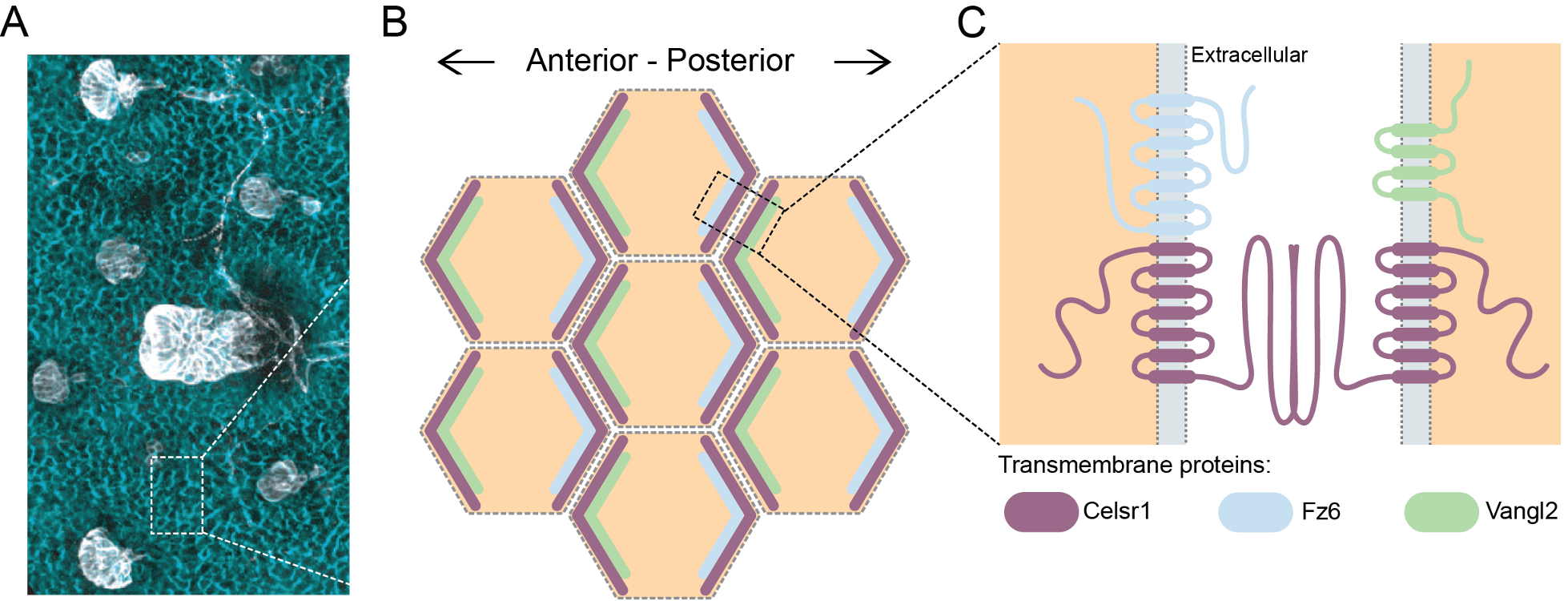
Tissue-level planar polarity is created at the cellular level by the asymmetric orientation of several core proteins, including Celsr1, Fz6, and Vangl2. These proteins are located on opposite sides of individual cells, with Vangl2 on the front side of the cell and Fz6 on the back (Figure 2). Celsr1 is present on both sides of the cell and forms a cell-cell junction, or a physical connection, linking two neighboring cells together and helping to maintain the asymmetric organization of Fz6 and Vangl2. These proteins and cell-cell junctions are present across vertebrates, including both humans and mice. Because of this, mice are a strong model to study these proteins and their role in larger development processes.
My dissertation research focused on a Celsr1 mutation identified in mice that results in NTDs.8 The mutation was nicknamed “Crash” or Crsh, as the mutant mice displayed defects in balance and frequently crashed into each other.8 At the protein level, the Crsh mutation prevents Celsr1 from properly interacting with itself, resulting in an unstable cell-cell junction.9 The working hypothesis is that under normal conditions, two Celsr1 proteins bind with each other on a membrane of a cell, a process called cis-dimerization, while two other Celsr1 proteins interact with that dimer across the gap between the cells to form a cell-cell junction, a process called trans-dimerization (Figure 3A). When Celsr1 harbors the Crsh mutation, the proteins can still interact across the junction, but are unable to cis-dimerize within the same cell, creating an unstable cell-cell junction and resulting in a loss of planar polarity (Figure 3B).

During cell division, Celsr1 is brought from the membrane into the cell in a process known as mitotic internalization.10 As it moves inward, Celsr1 pulls Fz6 and Vangl2 inside the cell as well, maintaining these proteins in a complex throughout cell division.10,11 Because this complex is held together very strongly, as Celsr1 is brought into the dividing cell, it also pulls in proteins from the membrane of the neighboring cell, a process called trans-endocytosis (Figure 3C).11 In effect, trans-endocytosis keeps the PCP proteins from both cells together during cell division.Previous research has shown that proper trans-endocytosis of these proteins is crucial for PCP during tissue development. However, what remains unknown is the role of Celsr1 cis-dimerization in controlling this internalization and how its disruption contributes to broader developmental defects.
My biggest question:
How does the Crsh mutation in Celsr1 alter mitotic internalization of PCP proteins?
While we know the Crsh mutation alters the ability of Celsr1 to cis-dimerize, it is unclear how this in turn impacts the internalization of PCP proteins during cell division. In my dissertation work I investigated the potential impact of the Crsh mutation on this mitotic internalization process. Because the Crsh mutation causes a less stable cell-cell junction, I hypothesized that it may disrupt proper mitotic internalization, particularly trans-endocytosis, because this process is heavily dependent on the Celsr1 complex forming a stable junction between two cells.
What I did to answer my question:
In my research, I used both mouse models and cell-based assays to study the internalization of different PCP proteins during cell division. First, I labeled Celsr1, Fz6, and Vangl2 in the back skin of both wildtype and Crsh mice and used an advanced microscope to visualize these proteins and determine if their mitotic internalization was disrupted by the Crsh mutation. Next, in cultured cell lines, I engineered the PCP proteins to have different colored tags in each cell, allowing me to determine which proteins are internalized from the membrane of the dividing cell and which proteins are internalized from neighboring cells. Finally, because the mutated Crsh protein is unable to cis-dimerize, I added an engineered protein fragment to the end of Crsh that allows me to treat with a drug that forces cis-dimerization to directly test the role of this dimerization in regulating PCP protein internalization.
What I found:
Using these methods, I determined that Celsr1 cis-dimerization is required for internalization of PCP proteins during mitosis. The Crsh mutation prevented Celsr1 and Vangl2 from being trans-endocytosed from the neighboring cell into the mitotic cell in both mouse models and cultured cells. Thus, because this mutant is unable to cis-dimerize, these results suggested that dimerization is necessary for proper Celsr1-mediated mitotic internalization. Interestingly, I also determined that Celsr1 cis-dimerization is required on both sides of the cell-cell junction for Celsr1 and Vangl2 trans-endocytosis to occur. This supports a model where the minimum functioning unit of Celsr1 is a structure of four proteins interacting: Celsr1 must be both cis-dimerized on each membrane and trans-dimerized across the cell-cell junction (Figure 2A). This model was further verified through rescue experiments using the engineered protein and dimerizing drug that rescues Crsh-induced defects in cis-dimerization. When I forced cis-dimerization of the Crsh protein through this method, PCP proteins were successfully trans-endocytosed from the neighboring cell. In other words, rescuing cis-dimerization of Celsr1 also rescued trans-endocytosis of the PCP proteins. These experiments solidified our conclusion that Celsr1 cis-dimerization is required on both sides of the cell-cell junction to mediate trans-endocytosis of PCP proteins.
These findings have further laid the groundwork for future research investigating how these proteins are moved between different compartments within cells after their internalization, and how mutations in Celsr1 may impact these movements. The PCP complex has previously been shown to be both degraded and recycled, with Celsr1 replaced back onto the membrane after the completion of cell division.10,11 Interestingly, preliminary work suggests that the Crsh mutation disrupts the balance between this recycling and degradation (unpublished data). Although the full implications of this alteration are unknown, it likely contributes to the broader developmental defects associated with the Crsh mutation, as shown for other mutations that disrupt proper movement of proteins between cellular compartments.12–17
Why are these findings important:
My findings reveal a broader mechanism of Celsr1 functioning and the specific protein-protein interactions needed to facilitate mitotic internalization, a process known to be crucial for proper planar polarity.10 Very little is known about mammalian PCP and how specific mutations result in developmental defects. While my dissertation work focused on one specific mutation in Celsr1, the mechanisms of PCP disruption identified by my findings are likely applicable across many mutations in the protein. These discoveries help us understand what specific protein processes are vital to directing critical developmental events, such as neural tube closure. Understanding how these mutations disrupt proper PCP protein functioning and lead to developmental deficiencies is crucial to relieving the health burden these mutations create.
If you would like to learn more about my thesis, you can find the peer-reviewed research in my published paper:
Latario SG, Saba T, Trejo B, Giang W, Zimmer SE, Devenport D, Stahley SN, Planar cell polarity complex internalization is dependent on Celsr1 cis-dimerization. iScience. 2026 Aug 21. DOI: 10.1016/j.isci.2026.116761
TL:DR
- Planar cell polarity (PCP) signaling regulates tissue patterning and has many crucial roles during development, including proper neural tube closure.
- Proper internalization of PCP proteins during mitosis is crucial to maintaining polarity during development.
- My dissertation work revealed that this mitotic internalization is mediated by Celsr1 cis-dimerization.
References
- Schneider, M.R., Schmidt-Ullrich, R., and Paus, R. (2009). The Hair Follicle as a Dynamic Miniorgan. Current Biology 19, R132–R142. https://doi.org/10.1016/j.cub.2008.12.005.
- Hu, M.S., Borrelli, M.R., Hong, W.X., Malhotra, S., Cheung, A.T.M., Ransom, R.C., Rennert, R.C., Morrison, S.D., Lorenz, H.P., and Longaker, M.T. (2018). Embryonic skin development and repair. Organogenesis 14, 46–63. https://doi.org/10.1080/15476278.2017.1421882.
- Phillips, H.M., Rhee, H.J., Murdoch, J.N., Hildreth, V., Peat, J.D., Anderson, R.H., Copp, A.J., Chaudhry, B., and Henderson, D.J. (2007). Disruption of Planar Cell Polarity Signaling Results in Congenital Heart Defects and Cardiomyopathy Attributable to Early Cardiomyocyte Disorganization. Circulation Research 101, 137–145. https://doi.org/10.1161/CIRCRESAHA.106.142406.
- Wu, G., Huang, X., Hua, Y., and Mu, D. (2011). Roles of planar cell polarity pathways in the development of neutral tube defects. J Biomed Sci 18, 66. https://doi.org/10.1186/1423-0127-18-66.
- Butler, M.T., and Wallingford, J.B. (2017). Planar cell polarity in development and disease. Nat Rev Mol Cell Biol 18, 375–388. https://doi.org/10.1038/nrm.2017.11.
- Wallingford, J.B., Niswander, L.A., Shaw, G.M., and Finnell, R.H. (2013). The continuing challenge of understanding, preventing, and treating neural tube defects. Science 339, 1222002. https://doi.org/10.1126/science.1222002.
- Cetera, M., Leybova, L., Woo, F.W., Deans, M., and Devenport, D. (2017). Planar cell polarity-dependent and independent functions in the emergence of tissue-scale hair follicle patterns. Developmental Biology 428, 188–203. https://doi.org/10.1016/j.ydbio.2017.06.003.
- Curtin, J.A., Quint, E., Tsipouri, V., Arkell, R.M., Cattanach, B., Copp, A.J., Henderson, D.J., Spurr, N., Stanier, P., Fisher, E.M., et al. (2003). Mutation of Celsr1 disrupts planar polarity of inner ear hair cells and causes severe neural tube defects in the mouse. Curr Biol 13, 1129–1133. https://doi.org/10.1016/s0960-9822(03)00374-9.
- Stahley, S.N., Basta, L.P., Sharan, R., and Devenport, D. (2021). Celsr1 adhesive interactions mediate the asymmetric organization of planar polarity complexes. eLife 10, e62097. https://doi.org/10.7554/eLife.62097.
- Devenport, D., Oristian, D., Heller, E., and Fuchs, E. (2011). Mitotic internalization of planar cell polarity proteins preserves tissue polarity. Nat Cell Biol 13, 893–902. https://doi.org/10.1038/ncb2284.
- Heck, B.W., and Devenport, D. (2017). Trans-endocytosis of Planar Cell Polarity Complexes during Cell Division. Current Biology 27, 3725-3733.e4. https://doi.org/10.1016/j.cub.2017.10.053.
- Giese, A.P., Ezan, J., Wang, L., Lasvaux, L., Lembo, F., Mazzocco, C., Richard, E., Reboul, J., Borg, J.-P., Kelley, M.W., et al. (2012). Gipc1 has a dual role in Vangl2 trafficking and hair bundle integrity in the inner ear. Development 139, 3775–3785. https://doi.org/10.1242/dev.074229.
- Merte, J., Jensen, D., Wright, K., Sarsfield, S., Wang, Y., Schekman, R., and Ginty, D.D. (2010). Sec24b selectively sorts Vangl2 to regulate planar cell polarity during neural tube closure. Nat Cell Biol 12, 41–46. https://doi.org/10.1038/ncb2002.
- Ossipova, O., Chuykin, I., Chu, C.-W., and Sokol, S.Y. (2015). Vangl2 cooperates with Rab11 and Myosin V to regulate apical constriction during vertebrate gastrulation. Development 142, 99–107. https://doi.org/10.1242/dev.111161.
- Ossipova, O., Kim, K., Lake, B.B., Itoh, K., Ioannou, A., and Sokol, S.Y. (2014). Role of Rab11 in planar cell polarity and apical constriction during vertebrate neural tube closure. Nat Commun 5, 3734. https://doi.org/10.1038/ncomms4734.
- Edwards, N.A., Rankin, S.A., Kashyap, A., Warren, A., Agricola, Z.N., Kenny, A.P., Kofron, M., Shen, Y., Chung, W.K., and Zorn, A.M. (2025). Disrupted endosomal trafficking of the Vangl-Celsr polarity complex underlies congenital anomalies in Xenopus trachea-esophageal morphogenesis. Developmental Cell 60, 2487-2502.e4. https://doi.org/10.1016/j.devcel.2025.04.026.
- Tower-Gilchrist, C., Zlatic, S.A., Yu, D., Chang, Q., Wu, H., Lin, X., Faundez, V., and Chen, P. (2019). Adaptor protein-3 complex is required for Vangl2 trafficking and planar cell polarity of the inner ear. MBoC 30, 2422–2434. https://doi.org/10.1091/mbc.E16-08-0592.
 Lions Talk Science